Ebola virus disease
From Wikipedia, the free encyclopedia
Jump to: navigation, search
"Ebola" redirects here. For other uses, see Ebola (disambiguation).

Ebola virus disease (EVD; also Ebola hemorrhagic fever (EHF)) or simply Ebola is a disease of humans and other primates caused by ebolaviruses. Signs and symptoms typically start between two days and three weeks after contracting the virus, with a fever, sore throat, muscle pain and headaches. Then, vomiting, diarrhea and rash usually follows, along with decreased function of the liver and kidneys. At this time, generally, some people begin to bleed both internally and externally. [1] Death, if it occurs, is typically six to sixteen days after symptoms appear and is often due to low blood pressure from fluid loss. [2]
The virus is acquired by contact with blood or other body fluids of an infected human or other animal. [1] This may also occur by direct contact with a recently contaminated item. [1] Spread through the air has not been documented in the natural environment. [3] Fruit bats are believed to be the normal carrier in nature, able to spread the virus without being affected. Humans become infected by contact with the bats or a living or dead animal that has been infected by bats. Once human infection occurs, the disease may spread between people as well. Male survivors may be able to transmit the disease via semen for nearly two months. To diagnose EVD, other diseases with similar symptoms such as malaria, cholera and other viral hemorrhagic fevers are first excluded. Blood samples are tested for viral antibodies, viral RNA, or the virus itself to confirm the diagnosis. [1]
Outbreak control requires a coordinated series of medical services, along with a certain level of community engagement. The necessary medical services include rapid detection and contact tracing, quick access to appropriate laboratory services, proper management of those who are infected, and proper disposal of the dead through cremation or burial. [1] [4] Prevention includes decreasing the spread of disease from infected animals to humans. [1] This may be done by only handling potentially infected bush meat while wearing protective clothing and by thoroughly cooking it before consumption. [1] It also includes wearing proper protective clothing and washing hands when around a person with the disease. [1] Samples of body fluids and tissues from people with the disease should be handled with special caution. [1]
No specific treatment for the disease is yet available. Efforts to help those who are infected are supportive and include giving either oral rehydration therapy (slightly sweetened and salty water to drink) or intravenous fluids. This supportive care improves outcomes. The disease has a high risk of death, killing between 25% and 90% of those infected with the virus (average is 50%). EVD was first identified in an area of Sudan (now part of South Sudan), as well as in Zaire (now the Democratic Republic of the Congo). The disease typically occurs in outbreaks in tropical regions of sub-Saharan Africa. [1] From 1976 (when it was first identified) through 2013, the World Health Organization reported a total of 1,716 cases. [1] [5] The largest outbreak to date is the ongoing 2014 West African Ebola outbreak, which is currently affecting Guinea, Sierra Leone, and Liberia. [6] [7] [8] As of 14 October 2014 [update], 9,216 suspected cases resulting in the deaths of 4,555 have been reported. [6] Efforts are under way to develop a vaccine; however, none yet exists. [1]
Contents [ hide]
Signs and symptoms

Signs and symptoms of Ebola. [9]
The time between exposure to the virus and the development of symptoms of the disease is usually 2 to 21 days. [1] [9] Estimates based on mathematical models predict that around 5% of cases may take greater than 21 days to develop. [10]
Symptoms usually begin with a sudden influenza-like stage characterized by feeling tired, fever, pain in the muscles and joints, headache, and sore throat. [1] [11] [12] The fever is usually greater than 38.3 °C (100.9 °F). [13] This is often followed by: vomiting, diarrhea and abdominal pain. [12] Shortness of breath and chest pain may occur next along with swelling, headaches and confusion. [12] In about half of cases the skin may develop a maculopapular rash (a flat red area covered with small bumps). [13]
In some cases, internal and external bleeding may occur. [1] This typically begins five to seven days after first symptoms. [14] All people show some decreased blood clotting. [13] Bleeding from mucous membranes or from sites of needle punctures is reported in 40–50% of cases. [15] This may result in the vomiting of blood, coughing up of blood or blood in stool. [16] Bleeding into the skin may create petechiae, purpura, ecchymoses, hematomas (especially around needle injection sites). [17] There may also be bleeding into the whites of the eyes. Heavy bleeding is uncommon and if it occurs is usually within the gastrointestinal tract. [13] [18]
Recovery may begin between 7 and 14 days after the start of symptoms. [12] Death, if it occurs, is typically 6 to 16 days from the start of symptoms and is often due to low blood pressure from fluid loss. [2] In general, the development of bleeding often indicates a worse outcome and this blood loss can result in death. [11] People are often in a coma near the end of life. [12] Those who survive often have ongoing muscle and joint pain, liver inflammation, and decreased hearing among other difficulties. [12]
Cause
Main articles: Ebolavirus (taxonomic group) and Ebola virus (specific virus)
Ebola virus disease in humans is caused by four of five viruses in the genus Ebolavirus. The four are Bundibugyo virus (BDBV), Sudan virus (SUDV), Taï Forest virus (TAFV), and one called, simply, Ebola virus (EBOV, formerly Zaire Ebola virus). [19] Ebola virus is the only member of the Zaire ebolavirus species and the most dangerous of the known EVD-causing viruses, as well as being responsible for the largest number of outbreaks. [20] The fifth virus, Reston virus (RESTV), is not thought to cause disease in humans, but has caused disease in other primates. [21] [22] These five viruses are closely related to marburgviruses. [19]
Transmission
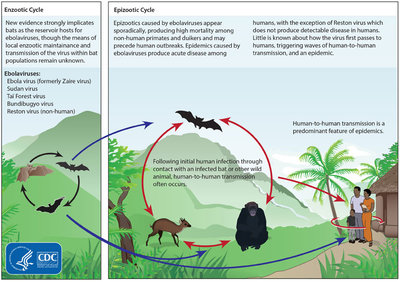
Life cycles of the Ebolavirus
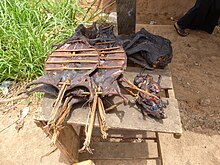
Bushmeat being prepared for cooking in Ghana, 2013. Human consumption of equatorial animals in Africa in the form of bushmeat has been linked to the transmission of diseases to people, including Ebola. [23]
The spread of Ebola between people occurs only by direct contact with the blood or body fluids of a person after symptoms have developed. [1] [3] Body fluids that may contain ebolaviruses include saliva, mucus, vomit, feces, sweat, tears, breast milk, urine, and semen. [24] Entry points include the nose, mouth, eyes, or open wounds, cuts and abrasions. [24] Contact with objects contaminated by the virus, particularly needles and syringes may also transmit the infection. [25] The virus is able to survive on objects for a few hours in a dried state and can survive for a few days within body fluids. [24] Ebola virus may be able to persist in the semen of survivors for up to seven weeks after recovery, which could give rise to infections via sexual intercourse. [1] Otherwise, people who have recovered are not infectious. [25] The potential for widespread infections in countries with medical systems capable of observing correct medical isolation procedures is considered low. [26] Usually when someone has symptoms, they are sufficiently unwell that they are unable to travel without assistance. [27]
Handling infected dead bodies is a risk, including embalming. [26] Because dead bodies are still infectious, traditional burial rituals may spread the disease. Nearly two thirds of the cases of Ebola infections in Guinea during the 2014 outbreak are believed to have been contracted via unprotected (or unsuitably protected) contact with infected corpses during certain Guinean burial rituals. [28] [29]
Healthcare workers treating those who are infected are at greatest risk of disease. [25] This occurs when they do not wear appropriate protective clothing such as masks, gowns, gloves and eye protection. [25] This is particularly common in parts of Africa where the health systems function poorly and where the disease mostly occurs. [30] Hospital-acquired transmission has also occurred in African countries due to the reuse of needles. [31] [32] Some healthcare centers caring for people with the disease do not have running water. [33] In the United States, spread has occurred due to inadequate isolation. [34]
While it is not entirely clear how Ebola initially spreads from animals to human, it is believed to involve direct contact with an infected wild animal or fruit bat. [25] In Africa wild animals, known as bushmeat, are hunted to eat. [35]
Airborne transmission has not been documented during EVD outbreaks. [3] Transmission among rhesus monkeys via breathable 0.8–1.2 µm aerosolized droplets has been demonstrated in the laboratory. [36] That airborne transmission does not appear to occur in humans may be due to there not being high enough levels of the virus in the lungs. [37] Spread by water or food other than bushmeat has also not been observed, [25] nor has spread by mosquitos or other insects. [25]
Reservoir Bats are considered the most likely natural reservoir of ebola virus. Plants, arthropods, and birds have also been considered. [1] [38] In the wild, transmission may occur when infected fruit bats drop partially eaten fruits or fruit pulp, then land mammals such as gorillas and duikers may feed on these fallen fruits. This chain of events forms a possible indirect means of transmission from the natural host species to other animal species, which has led to research into viral shedding in the saliva of fruit bats. Fruit production, animal behavior, and other factors vary at different times and places that may trigger outbreaks among animal populations. [39]
Bats were known to reside in the cotton factory in which the first cases of the 1976 and 1979 outbreaks were observed, and they have also been implicated in Marburg virus infections in 1975 and 1980. [40] Of 24 plant species and 19 vertebrate species experimentally inoculated with EBOV, only bats became infected. [41] The bats displayed no clinical signs and is evidence that these bats are a reservoir species of the virus. In a 2002–2003 survey of 1,030 animals including 679 bats from Gabon and the Republic of the Congo, 13 fruit bats were found to contain EBOV RNA fragments. [42] As of 2005, three types of fruit bats ( Hypsignathus monstrosus, Epomops franqueti, and Myonycteris torquata) have been identified as being in contact with EBOV. They are now suspected to represent the EBOV reservoir hosts. [43] [44] Antibodies against Zaire and Reston viruses have been found in fruit bats in Bangladesh, thus identifying potential virus hosts and signs of the filoviruses in Asia. [45]
Between 1976 and 1998, in 30,000 mammals, birds, reptiles, amphibians and arthropods sampled from outbreak regions, no Ebola virus was detected apart from some genetic traces found in six rodents ( Mus setulosus and Praomys) and one shrew ( Sylvisorex ollula) collected from the Central African Republic. [40] [46] Traces of EBOV were detected in the carcasses of gorillas and chimpanzees during outbreaks in 2001 and 2003, which later became the source of human infections. However, the high lethality from infection in these species makes them unlikely as a natural reservoir. [40]
Virology
Main articles: Ebolavirus (taxonomic group) and Ebola virus (specific virus)

Electron micrograph of an Ebola virus virion
They contain single-strand, non-infectious RNA genomes. [47] Ebolavirus genomes are approximately 19 kilobase pairs long and contain seven genes in the order 3'-UTR-NP-VP35-VP40-GP-VP30-VP24-L- 5'-UTR. [48] The genomes of the five different ebolaviruses (BDBV, EBOV, RESTV, SUDV, and TAFV) differ in sequence and the number and location of gene overlaps. Like all filoviruses, ebolavirions are filamentous particles that may appear in the shape of a shepherd's crook or in the shape of a "U" or a "6", and they may be coiled, toroid, or branched. [48] In general, ebolavirions are 80 nm in width, but vary somewhat in length. In general, the median particle length of ebolaviruses ranges from 974 to 1,086 nm (in contrast to marburgvirions, whose median particle length was measured at 795–828 nm), but particles as long as 14,000 nm have been detected in tissue culture. [49]
Their life cycle begins with virion attachment to specific cell-surface receptors, followed by fusion of the virion envelope with cellular membranes and the concomitant release of the virus nucleocapsid into the cytosol. Ebolavirus' structural glycoprotein (known as GP1,2) is responsible for the virus' ability to bind to and infect targeted cells. [50] The viral RNA polymerase, encoded by the L gene, partially uncoats the nucleocapsid and transcribes the genes into positive-strand mRNAs, which are then translated into structural and nonstructural proteins. The most abundant protein produced is the nucleoprotein, whose concentration in the cell determines when L switches from gene transcription to genome replication. Replication results in full-length, positive-strand antigenomes that are, in turn, transcribed into negative-strand virus progeny genome copy. Newly synthesized structural proteins and genomes self-assemble and accumulate near the inside of the cell membrane. Virions bud off from the cell, gaining their envelopes from the cellular membrane they bud from. The mature progeny particles then infect other cells to repeat the cycle. The Ebola virus genetics are difficult to study due to its virulent nature. [51]
Pathophysiology
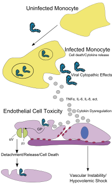
Pathogenesis schematic
Cells lining the inside of blood vessels ( endothelial cells), macrophages, monocytes, and liver cells are the main targets of infection. After infection, a secreted glycoprotein, known as small soluble glycoprotein (sGP) or as the Ebola virus glycoprotein (GP), is synthesized. Ebolavirus replication overwhelms protein synthesis of infected cells and host immune defenses. The GP forms a trimeric complex, which binds the virus to the endothelial cells. The sGP forms a dimeric protein that interferes with the signaling of neutrophils, a type of white blood cell, which allows the virus to evade the immune system by inhibiting early steps of neutrophil activation. These white blood cells also serve as carriers to transport the virus throughout the entire body to places such as the lymph nodes, liver, lungs, and spleen. [52] The presence of viral particles and cell damage resulting from viruses budding out of the cell causes the release of chemical signals (such as TNF-a, IL-6, and IL-8), which are molecular signals for fever and inflammation. The damage to human cells, caused by infection of the endothelial cells, decreases blood vessel integrity. This loss of vascular integrity is furthered with the synthesis of GP, which reduces specific integrins responsible for cell adhesion to the intercellular structure, and damage to the liver, which leads to improper clotting. [53]
Filoviral infection is also known to interfere with proper functioning of the innate immune system. [54] Ebolavirus proteins have demonstrated the ability to blunt the human immune system's response to viral infections by interfering with cells' ability to produce and respond to interferon proteins such as interferon-alpha, interferon-beta, and interferon gamma. [50] [55] This interference is accomplished by the VP24 and VP35 ebolavirus structural proteins. When cells are infected with ebolavirus, receptors located in the cell's cytosol (such as RIG-I and MDA5) or outside of the cytosol (such as Toll-like receptor 3, Toll-like receptor 7, Toll-like receptor 8, and Toll-like receptor 9), recognize infectious molecules associated with the virus. [50] After these receptors are activated, proteins including interferon regulatory factor 3 and interferon regulatory factor 7 start a signaling cascade that leads to the expression of type 1 interferons. [50] Type 1 interferons are then released and bind to neighboring uninfected cells expressing the IFNAR1 and IFNAR2 receptors on their surface. [50] Once interferon has bound to its receptors on the neighboring uninfected cell, the signaling proteins STAT1 and STAT2 are activated and move to the uninfected cell's nucleus. [50] This triggers the expression of interferon-stimulated genes, which code for proteins that have antiviral properties. [50] Ebolavirus' V24 protein prevents the STAT1 signaling protein in the neighboring uninfected cells from entering the cell's nucleus and therefore prevents the creation of these antiviral proteins. [50] A separate ebolavirus protein, known as VP35, directly inhibits the production of interferon-beta. [55]
DiagnosisWhen the diagnosis of EVD is suspected, the travel and work history along with exposure to wildlife are important factors to consider. The diagnosis is confirmed by isolating the virus, detecting its RNA or proteins, or detecting antibodies against the virus in a person's blood. Isolating the virus by cell culture, detecting the viral RNA by polymerase chain reaction (PCR) and detecting proteins by enzyme-linked immunosorbent assay (ELISA) works best early and in those who have died from the disease. Detecting antibodies against the virus works best late in the disease and in those who recover. [56]
During an outbreak, virus isolation is often not feasible. The most common diagnostic methods are therefore real-time PCR and ELISA detection of proteins, which can be performed in field or mobile hospitals. [57] Filovirions can be seen and identified in cell culture by electron microscopy due to their unique filamentous shapes, but electron microscopy cannot tell the difference between the various filoviruses despite there being some length differences. [49]
Laboratory testingChanges on laboratory tests as a result of Ebola virus disease include a low platelet count in the blood, an initially decreased white blood cell count followed by an increase in the white blood cell count, elevated levels of the liver enzymes alanine aminotransferase (ALT) and aspartate aminotransferase (AST), and abnormalities in clotting often consistent with disseminated intravascular coagulation (DIC) such as a prolonged prothrombin time, partial thromboplastin time, and bleeding time. [58]
Differential diagnosisEarly symptoms of EVD may be similar to those of other diseases common in Africa including malaria and dengue fever. [11] The symptoms are also similar to those of Marburg virus disease and other viral hemorrhagic fevers. [59]
The complete differential diagnosis is long and includes many other infectious diseases such as typhoid fever, shigellosis, rickettsial diseases, cholera, sepsis, borreliosis, EHEC enteritis, leptospirosis, scrub typhus, plague, Q fever, candidiasis, histoplasmosis, trypanosomiasis, visceral leishmaniasis, measles, and viral hepatitis among others. [60] Non-infectious diseases that can be confused with EVD include acute promyelocytic leukemia, hemolytic uremic syndrome, snake envenomation, clotting factor deficiencies/platelet disorders, thrombotic thrombocytopenic purpura, hereditary hemorrhagic telangiectasia, Kawasaki disease, and warfarin poisoning among others. [61] [62] [63] [64]
Prevention

A researcher working with the Ebola virus while wearing a BSL-4 positive pressure suit to avoid infection
Infection controlRecommended measures for people caring for those infected with Ebola include the wearing of protective clothing including masks, gloves, gowns, and goggles. [65] These same measures are recommended for those who may handle objects contaminated by the infected person's body fluids. [66] The infected person should be barrier-isolation from other people. [65] All equipment, medical waste, patient waste, and surfaces that may have come into contact with body fluids require disinfection. [66] Education on the proper suit-up and removal of personal protective equipment is also required. In Sierra Leone, the typical training period for the use of such safety equipment lasts approximately 12 days. [67]
During the 2014 outbreak kits were put together to help familes treat Ebola in their homes which includes protective clothing as well as chlorine powder and other cleaning supplies. [68] Education of those who provide care in these techniques, and the provision of such barrier-separation supplies has been a priority of the Doctors Without Borders organization. [69]
Ebolaviruses can be eliminated with heat (heating for 30 to 60 minutes at 60 °C or boiling for 5 minutes). To disinfect surfaces, some lipid solvents such as some alcohol-based products, detergents, sodium hypochlorite (bleach) or calcium hypochlorite (bleaching powder), and other suitable disinfectants at appropriate concentrations can be used. [70] [71]
Education of the general public of the risk factors for Ebola infection and of the protective measures individuals can take is recommended by the World Health Organization. [1] These measures include avoiding direct contact with infected people and regular hand washing using soap and water. [72]
Bushmeat, an important source of protein in the diet of some Africans, should be handled with appropriate protective clothing and thoroughly cooked before consumption. [1] Some research suggests that an outbreak in the wild animals used for consumption may result in a corresponding human outbreak. Since 2003, such animal outbreaks have been monitored with the aim of predicting and preventing Ebola outbreaks in humans. [73]
If a person with Ebola dies, direct contact with the body should be avoided. [65] Certain burial rituals, which might have included making any kind of direct contact with a dead body, require reformulation such that they consistently maintain a proper protective barrier between the bead body and the living. [74] [75] It is recommended that the bodies of people who have died from Ebola be buried or cremated only with proper care. [76] Social anthropologists may help find alternatives to traditional rules for burials. [77]
Transportation crews are instructed to follow a certain isolation procedure should anyone exhibit symptoms resembling the Ebola virus disease. [78] The World Health Organization as of Aug 14, 2014 does not consider travel bans to be useful in decreasing spread. [27]
In laboratories where diagnostic testing is carried out, biosafety level 4-equivalent containment is required, since ebolaviruses are World Health Organization Risk Group 4 pathogens. Laboratory researchers must be properly trained in BSL-4 practices and wear proper personal protective equipment.
Quarantine Quarantine, also known as enforced isolation, is usually effective in decreasing spread. [79] [80] Governments often quarantine areas where the disease is occurring or individuals who may transmit the disease outside of an initial area. [81] In the United States, the law allows quarantine of those infected with ebolaviruses. [82] During the 2014 outbreak, Liberia closed schools. [83] On October 16, 2014, some schools were closed in Ohio and Texas as a precaution after one of two nurses who contracted Ebola (after caring for a person with Ebola) returned to the Cleveland area and may have been on the same plane as some students, teachers, and parents of students from those schools. [84]
Contact tracing Contact tracing is regarded as important to contain an outbreak. It involves finding everyone who had close contact with infected individuals and watching for signs of illness for 21 days. If any of these contacts comes down with the disease, they should be isolated, tested, and treated. Then repeat the process by tracing the contacts' contacts. [85] [86]
TreatmentStandard support
A hospital isolation ward in Gulu, Uganda, during the October 2000 outbreak
No ebolavirus-specific treatment is currently approved. [87] However, survival is improved by early supportive care with rehydration and symptomatic treatment. [1] Treatment is primarily supportive in nature. [88] These measures may include management of pain, nausea, fever and anxiety, as well as rehydration via the oral or by intravenous route. [88] Blood products such as packed red blood cells, platelets or fresh frozen plasma may also be used. [88] Other regulators of coagulation have also been tried including heparin in an effort to prevent disseminated intravascular coagulation and clotting factors to decrease bleeding. [88] Antimalarial medications and antibiotics are often used before the diagnosis is confirmed, [88] though there is no evidence to suggest such treatment is in any way helpful.
Intensive care Intensive care is often used in the developed world. [17] This may include maintaining blood volume and electrolytes (salts) balance as well as treating any bacterial infections that may develop. [17] Dialysis may be needed for kidney failure while extracorporeal membrane oxygenation may be used for lung dysfunction. [17]
Alternative medicineThe Food and Drug Administration (FDA) advises people to be careful of advertisements making unverified or fraudulent claims of benefits supposedly gained from various anti-Ebola products. [89] The FDA has already sent out at least one letter of warning to a seller of colloidal silver who made unverified claims of Ebola related benefits, supposedly derived from the use of their products. [90]
PrognosisEbola virus disease has a high risk of death in those infected which varies between 25 percent and 90 percent of those infected. [1] [91] As of September 2014 [update], the average risk of death among those infected is 50%. [1] The risk of death was 90% in the 2002–2003 Republic of the Congo outbreak. [92] There are indications based on variations between countries that early and effective treatment of symptoms (e.g., supportive care to prevent dehydration) may reduce the risk of death. [93]
If an infected person survives, recovery may be quick and complete. Prolonged cases are often complicated by the occurrence of long-term problems, such as inflammation of the testicles, joint pains, muscle pains, skin peeling, or hair loss. Eye symptoms, such as light sensitivity, excess tearing, iritis, iridocyclitis, choroiditis, and blindness have also been described.
Epidemiology

Cases of ebola fever in Africa from 1979 to 2008.
For more about specific outbreaks and their descriptions, see List of Ebola outbreaks.

CDC worker incinerates medical waste from Ebola patients in Zaire in 1976
The disease typically occurs in outbreaks in tropical regions of Sub-Saharan Africa. [1] From 1976 (when it was first identified) through 2013, the World Health Organization reported 1,716 confirmed cases. [1] [5] The largest outbreak to date is the ongoing 2014 West Africa Ebola virus outbreak, which is affecting Guinea, Sierra Leone, Liberia and Nigeria. [7] [8] As of 15 October, 8,997 suspected cases have been identified, with 4,496 deaths. [94]
1976Sudan outbreakThe first known outbreak of Ebola virus disease (EVD) was identified only after the fact, occurring between June and November 1976 in Nzara, South Sudan, [19] [95] (then part of Sudan) and was caused by Sudan virus (SUDV). The Sudan outbreak infected 284 people and killed 151. The first identifiable case in Sudan occurred on 27 June in a storekeeper in a cotton factory in Nzara, who was hospitalized on 30 June and died on 6 July. [96] [97] While the WHO medical staff involved in the Sudan outbreak were aware that they were dealing with a heretofore unknown disease, the actual "positive identification" process and the naming of the virus did not occur until some months later in the Democratic Republic of the Congo. [96]
Zaire outbreak
See also: Yambuku § Ebola outbreak
On 26 August 1976, a second outbreak of EVD began in Yambuku, Zaire, a small rural village in Mongala District in northern Democratic Republic of the Congo (then known as Zaire). [98] [99] This outbreak was caused by Ebola virus (EBOV), formerly designated Zaire ebolavirus, which is a different member of the genus Ebolavirus than in the first Sudan outbreak. The first person infected with the disease was village school headmaster Mabalo Lokela, who began displaying symptoms on August 26, 1976. [100] Lokela had returned from a trip to Northern Zaire near the Central African Republic border, having visited the Ebola River between 12–22 August. He was originally believed to have malaria and was given quinine. However, his symptoms continued to worsen, and he was admitted to Yambuku Mission Hospital on September 5. Lokela died on September 8, fourteen days after he began displaying symptoms. [101] [102] [103] [104]
Soon after Lokela's death, others who had been in contact with him also died, and people in the village of Yambuku began to panic. This led the country's Minister of Health along with Zaire President Mobutu Sese Seko to declare the entire region, including Yambuku and the country's capital, Kinshasa, a quarantine zone. No one was permitted to enter or leave the area, with roads, waterways, and airfields placed under martial law. Schools, businesses and social organizations were closed. [105] Researchers from the CDC, including Peter Piot, co-discoverer of Ebola, later arrived to assess the effects of the outbreak, observing that "the whole region was in panic." [106] The outbreak lasted 26 days, with the quarantine lasting two weeks. Among the reasons that researchers speculated caused the disease to disappear, were the precautions taken by locals, the quarantine of the area, and possibly most important, the discontinuance of reusing needles by local nurses. [105]
During this outbreak, Dr. Ngoy Mushola recorded the first clinical description of Ebola virus disease in Yambuku, where he wrote the following in his daily log: "The illness is characterized with a high temperature of about 39°C, hematemesis, diarrhea with blood, retrosternal abdominal pain, prostration with "heavy" articulations, and rapid evolution death after a mean of three days." [107]
The virus responsible for the initial outbreak, first thought to be Marburg virus, was later identified as a new type of virus related to marburgviruses. Virus strain samples isolated from both outbreaks were named as the "Ebola virus" after the Ebola River, located near the originally identified viral outbreak site in Zaire. [108] Reports conflict about who initially coined the name: either Karl Johnson of the American CDC team [109] or Belgian researchers. [110] Subsequently a number of other cases were reported, almost all centered on the Yambuku mission hospital or having close contact with another case. [100] 318 cases and 280 deaths (a 88% fatality rate) occurred in the DRC. [111] Although it was assumed that the two outbreaks were connected, scientists later realized that they were caused by two distinct ebolaviruses, SUDV and EBOV. [99] The Zaire outbreak was contained with the help of the World Health Organization and transport from the Congolese air force, by quarantining villagers, sterilizing medical equipment, and providing protective clothing.
1995 to 2013The second major outbreak occurred in 1995 in the Democratic Republic of Congo, affecting 315 and killing 254. The next major outbreak occurred in Uganda in 2000, affecting 425 and killing 224; in this case the Sudan virus was found to be the ebolavirus species responsible for the outbreak. [112] In 2003 there was an outbreak in the Republic of Congo that affected 143 and killed 128, a death rate of 90%, the highest to date. [113]
In 2004 a Russian scientist died from Ebola after sticking herself with an infected needle. [114]
In August 2007, 103 people were infected by a suspected hemorrhagic fever outbreak in the village of Kampungu, Democratic Republic of the Congo. The outbreak started after the funerals of two village chiefs, and 217 people in four villages fell ill. [112] [115] [116] The 2007 outbreak eventually affected 264 individuals and resulted in the deaths of 187. [1]
On 30 November 2007, the Uganda Ministry of Health confirmed an outbreak of Ebola in the Bundibugyo District in Western Uganda. After confirmation of samples tested by the United States National Reference Laboratories and the Centers for Disease Control, the World Health Organization confirmed the presence of a new species of Ebolavirus, which was tentatively named Bundibugyo. [117] The WHO reported 149 cases of this new strain and 37 of those led to deaths. [1]
The WHO confirmed two small outbreaks in Uganda in 2012. The first outbreak affected 7 people and resulted in the death of 4 and the second affected 24, resulting in the death of 17. The Sudan variant was responsible for both outbreaks. [1]
On 17 August 2012, the Ministry of Health of the Democratic Republic of the Congo reported an outbreak of the Ebola-Bundibugyo variant [118] in the eastern region. [119] [120] Other than its discovery in 2007, this was the only time that this variant has been identified as the ebolavirus responsible for an outbreak. The WHO revealed that the virus had sickened 57 people and claimed 29 lives. The probable cause of the outbreak was tainted bush meat hunted by local villagers around the towns of Isiro and Viadana. [1] [121]
2014 West African outbreak
Main article: Ebola virus epidemic in West Africa
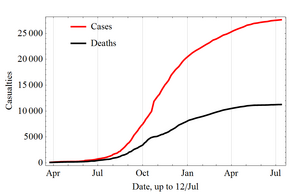
Increase over time in the cases and deaths during the 2014 outbreak
In March 2014, the World Health Organization (WHO) reported a major Ebola outbreak in Guinea, a western African nation. [122] Researchers traced the outbreak to a two-year old child who died on 28 December 2013. [123] [124] The disease then rapidly spread to the neighboring countries of Liberia and Sierra Leone. It is the largest Ebola outbreak ever documented, and the first recorded in the region. [122]
On 8 August 2014, the WHO declared the epidemic to be an international public health emergency. Urging the world to offer aid to the affected regions, the Director-General said, "Countries affected to date simply do not have the capacity to manage an outbreak of this size and complexity on their own. I urge the international community to provide this support on the most urgent basis possible." [125] By mid-August 2014, Doctors Without Borders reported the situation in Liberia's capital Monrovia as "catastrophic" and "deteriorating daily". They reported that fears of Ebola among staff members and patients had shut down much of the city’s health system, leaving many people without treatment for other conditions. [126] By late August 2014, the disease had spread to Nigeria, and one case was reported in Senegal. [127] [128] [129] [130] On 30 September 2014, the first confirmed case of Ebola in the United States was diagnosed. [131] The patient died eight days later. [132]
Aside from the human cost, the outbreak has severely eroded the economies of the affected countries. A Financial Times report suggested the economic impact of the outbreak could kill more people than the virus itself. As of 23 September, in the three hardest hit countries, Liberia, Sierra Leone, and Guinea, there were only 893 treatment beds available while the current need was 2122. In a 26 September statement, the WHO said, "The Ebola epidemic ravaging parts of West Africa is the most severe acute public health emergency seen in modern times. Never before in recorded history has a biosafety level four pathogen infected so many people so quickly, over such a broad geographical area, for so long." [133]
As of 14 October 2014 [update], 9,216 suspected cases and 4,555 deaths had been reported; [6] [94] however, the World Health Organization has said that these numbers may be vastly underestimated. [134] The WHO reports that more than 216 healthcare workers are among the dead, partly due to the lack of equipment and long hours. [135] [136]
2014 international spread
See also: Ebola virus cases in the United States
As of 15 October 2014, there have been 17 cases of Ebola treated outside of Africa, four of whom have died. [137] In early October, Teresa Romero, a 44-year-old Spanish nurse, contracted Ebola after caring for a priest who had been repatriated from west Africa. This was the first transmission of the virus to occur outside of Africa. [138]
On 19 September, Eric Duncan flew from his native Liberia to Texas; five days later he began showing symptoms and visited a hospital, but was sent home. His condition worsened and he returned to the hospital on 28 September, where he passed away on 8 October. [139] Health officials confirmed a diagnosis of Ebola on 30 September—the first case in the United States. [34] On 12 October, the CDC confirmed that a nurse in Texas who had treated Duncan was found to be positive for the Ebola virus, the first known case of the disease to be contracted in the United States. [140] On 15 October a second Texas healthcare worker was confirmed to have the virus. [141]
Society and cultureEbolavirus is classified as a biosafety level 4 agent, as well as a Category A bioterrorism agent by the Centers for Disease Control and Prevention. It has the potential to be weaponized for use in biological warfare, [142] [143] and was investigated by the Biopreparat for such use, but might be difficult to prepare as a weapon of mass destruction because the virus becomes ineffective quickly in open air. [144]
Literature Richard Preston's 1995 best-selling book, The Hot Zone, dramatized the Ebola outbreak in Reston, Virginia. [145]
William Close's 1995 Ebola: A Documentary Novel of Its First Explosion and 2002 Ebola: Through the Eyes of the People focused on individuals' reactions to the 1976 Ebola outbreak in Zaire. [146]
Tom Clancy's 1996 novel, Executive Orders, involves a Middle Eastern terrorist attack on the United States using an airborne form of a deadly Ebola virus strain named "Ebola Mayinga" (see Mayinga N'Seka). [147]
Other animalsWild animalsEbola has a high mortality among primates. [148] Frequent outbreaks of Ebola may have resulted in the deaths of 5,000 gorillas. [149] Outbreaks of Ebola may have been responsible for an 88% decline in tracking indices of observed chimpanzee populations in 420 square kilometer Lossi Sanctuary between 2002 and 2003. [150] Transmission among chimpanzees through meat consumption constitutes a significant risk factor, while contact between the animals, such as touching dead bodies and grooming, is not. [151]
Recovered carcasses from gorillas contain multiple Ebola virus strains, which suggest multiple introductions of the virus. Bodies decompose quickly and carcasses are not infectious after three to four days. Contact between gorilla groups is rare, suggesting transmission among gorilla groups is unlikely, and that outbreaks result from transmission between viral reservoir and animal populations. [150]
Domesticated animals
For more about the outbreak in Virginia, US, see Reston virus.
In late 1989, Hazelton Research Products' Reston Quarantine Unit in Reston, Virginia, suffered a mysterious outbreak of fatal illness amongst certain lab monkeys. This lab outbreak was initially diagnosed as Simian hemorrhagic fever virus (SHFV), and occurred amongst a shipment of crab-eating macaque monkeys imported from the Philippines.
Hazelton's veterinary pathologist sent tissue samples from dead animals to the United States Army Medical Research Institute of Infectious Diseases (USAMRIID) at Fort Detrick, Maryland, where an ELISA test indicated the antibodies present in the tissue were a response to ebola virus and not SHFV. [152] An electron microscopist from USAMRIID discovered filoviruses similar in appearance to Ebola in the tissue samples sent from Hazelton Research Products' Reston Quarantine Unit. [153]
Shortly afterward, a US Army team headquartered at USAMRIID went into action to euthanize the monkeys which had not yet died, bringing those monkeys and those which had already died of the disease to Ft. Detrick for study by the Army's veterinary pathologists and virologists, and eventual disposal under safe conditions. [152]
Blood samples were taken from 178 animal handlers during the incident. [154] Of those, six animal handlers eventually seroconverted, including one who had cut himself with a bloody scalpel. [52] [155] Despite its status as a Level-4 organism and its apparent pathogenicity in monkeys, when the handlers did not become ill, the CDC concluded that the virus had a very low pathogenicity to humans. [155] [156]
The Philippines and the United States had no previous cases of Ebola infection, and upon further isolation, researchers concluded it was another strain of Ebola, or a new filovirus of Asian origin, which they named Reston ebolavirus (RESTV) after the location of the incident. [152]
Reston virus (RESTV) can be transmitted to pigs. [157]
Since the initial outbreak it has since been found in nonhuman primates in Pennsylvania, Texas, and Italy, [81] where the virus had infected pigs. [158]
In 2012 it was demonstrated that the virus can travel without contact from pigs to nonhuman primates, although the same study failed to achieve transmission in that manner between primates. [157] [159] According to the WHO, routine cleaning and disinfection of pig (or monkey) farms with sodium hypochlorite or other detergents should be effective in inactivating the Reston ebolavirus. If an outbreak is suspected, the area must be immediately quarantined. [1]
While pigs that have been infected with RESTV tend to show symptoms of the disease, it has been shown that dogs may become infected with EBOV and remain asymptomatic. Dogs in some parts of Africa scavenge for their food and it is known that they sometimes eat infected animals and the corpses of humans. Although they remain asymptomatic, a 2005 survey of dogs during an EBOV outbreak found that over 31.8% showed a seroprevalence for EBOV closest to an outbreak versus 9% a farther distance away. [160]
ResearchA number of experimental treatments are being studied. [161] In the United States, the Food and Drug Administration (FDA)'s animal efficacy rule is being used to demonstrate reasonable safety to obtain permission to treat people who are infected with Ebola. It is being used because the normal path for testing drugs is not possible for diseases caused by dangerous pathogens or toxins. Experimental drugs are made available for use with the approval of regulatory agencies under named patient programs, known in the US as "expanded access". [162] On 12 August 2014 the WHO released a statement that the use of not yet proven treatments is ethical in certain situations in an effort to treat or prevent the disease. [163]
Medications

Researchers looking at slides of cultures of cells that make monoclonal antibodies. These are grown in a lab and the researchers are analyzing the products to select the most promising.
AntiviralsA number of antiviral medications are being studied.
- Favipiravir, an anti-viral drug approved in Japan for stockpiling against influenza pandemics, appears to be useful in a mouse model of Ebola. [11] [164] On 4 October 2014, it was reported that a French nun who contracted Ebola while volunteering in Liberia was cured with Favipiravir treatment. [165]
- BCX4430 is a broad-spectrum small molecule antiviral drug developed by BioCryst Pharmaceuticals and undergoing animal testing as a potential human treatment for Ebola by USAMRIID. [166] The drug has been approved to progress to Phase 1 trials, expected late in 2014. [167]
- Brincidofovir is a broad-spectrum antiviral drug. Its maker has been granted FDA approval to proceed with a trial to test its safety and effectiveness in Ebola patients. [168] It has been used to treat the first patient diagnosed with Ebola in the USA, after he had recently returned from Liberia. [169] [170]
- Lamivudine, an antiviral drug which is usually used to treat HIV / AIDS, was reported in September 2014 to have been used successfully to treat 13 out of 15 Ebola-infected patients by a doctor in Liberia, as part of a combination therapy also involving intravenous fluids and antibiotics to combat opportunistic bacterial infection of Ebola-compromised internal organs. [171] Western virologists have however expressed caution about the results, due to the small number of patients treated and confounding factors present. Researchers at the NIH stated that lamivudine had so far failed to demonstrate anti-Ebola activity in preliminary in vitro tests, but that they would continue to test it under different conditions and would progress it to trials if even slight evidence for efficacy is found. [172]
- JK-05 is an antiviral drug developed by the Chinese company Sihuan Pharmaceutical along with the Chinese Academy of Military Medical Sciences. It is reportedly being fast tracked through human trials for Ebola treatment after successful tests in mice. [173] [174]
- Lack of available treatment options has spurred research into a number of other possible antivirals targeted against Ebola, including natural products such as scytovirin and griffithsin, [175] [176] as well as synthetic drugs including FGI-103, FGI-104, FGI-106, dUY11 and LJ-001, [177] and other newer agents. [178] [179] [180]
Antisense technologyOther promising treatments rely on antisense technology. Both small interfering RNAs (siRNAs) and phosphorodiamidate morpholino oligomers (PMOs) targeting Ebola virus (EBOV) RNA polymerase L protein may prevent disease in nonhuman primates. [181] [182] TKM-Ebola is a small interfering RNA compound, currently being tested in a Phase I clinical trial in humans. [183] [184] Sarepta Therapeutics has completed a Phase I clinical trial with its PMO protecting up to 80-100 percent of the non-human primates tested. [185]
Antibodies ZMapp is a monoclonal antibody vaccine. The limited supply of the drug has been used to treat a small number of individuals infected with the Ebola virus. Although some individuals have recovered, the outcome is not considered statistically significant. [186] ZMapp has proved effective in a trial involving Rhesus macaque monkeys. [183] [187]
Researchers in Thailand claim to have developed an antibody-based treatment for Ebola using synthesized fragments of the virus. It has not been tested against Ebola itself. Scientists from the WHO and NIH have offered to test the treatment against live Ebola virus, but there is still a great deal of development needed before human trials. [188]
OtherTwo selective estrogen receptor modulators usually used to treat infertility and breast cancer ( clomiphene and toremifene) have been found to inhibit the progress of Ebola virus in vitro as well as in infected mice. Ninety percent of the mice treated with clomiphene and fifty percent of those treated with toremifene survived the tests. [189] The study authors conclude that given their oral availability and history of human use, these drugs would be candidates for treating Ebola virus infection in remote geographical locations, either on their own or together with other antiviral drugs.
A 2014 study found that three ion channel blockers used in the treatment of heart arrhythmias, amiodarone, dronedarone and verapamil, block the entry of Ebola virus into cells in vitro. [190]
Melatonin has also been suggested as a potential treatment for Ebola based on promising in vitro results. [191]
Blood productsThe WHO has stated that transfusion of whole blood or purified serum from Ebola survivors is the therapy with the greatest potential to be implemented immediately, although there is little information as to its efficacy. [192] September 2014, WHO issued an interim guideline for this therapy. [193] The blood serum from those who have survived an infection is currently being studied to see if it is an effective treatment. [194] During a meeting arranged by WHO this research was deemed to be a top priority. [194] Seven of eight people with Ebola survived after receiving a transfusion of blood donated by individuals who had previously survived the infection in an 1999 outbreak in the Democratic Republic of the Congo. [88] [195] This treatment, however, was started late in the disease meaning they may have already been recovering on their own and the rest of their care was better than usual. [88] Thus this potential treatment remains controversial. [17] Intravenous antibodies appear to be protective in non-human primates who have been exposed to large doses of Ebola. [196] The World Health Organization has approved the use of convalescent serum and whole blood products to treat people with Ebola. [197]
VaccineAs of September 2014, no vaccine was approved by the United States Food and Drug Administration (FDA) for clinical use in humans. [194] [198] It was hoped that one would be initially available by November 2014. [194] The most promising candidates are DNA vaccines [199] or vaccines derived from adenoviruses, [200] vesicular stomatitis Indiana virus (VSIV) [201] [202] [203] or filovirus-like particles (VLPs) [204] because these candidates could protect nonhuman primates from ebolavirus-induced disease. DNA vaccines, adenovirus-based vaccines, and VSIV-based vaccines have entered clinical trials. [205] [206] [207] [208]
Vaccines have protected nonhuman primates. Immunization takes six months, which impedes the counter-epidemic use of the vaccines. Searching for a quicker onset of effectiveness, in 2003, a vaccine using an adenoviral (ADV) vector carrying the Ebola spike protein was tested on crab-eating macaques. Twenty-eight days later, they were challenged with the virus and remained resistant. [200] A vaccine based on attenuated recombinant vesicular stomatitis virus (VSV) vector carrying either the Ebola glycoprotein or the Marburg glycoprotein in 2005 protected nonhuman primates, [209] opening clinical trials in humans. [205] Over a three month human trial, three vaccinations safely induced an immune response. Individuals for a year were followed, and, in 2006, a study testing a faster-acting, single-shot vaccine began; this new study was completed in 2008. [206] Trying the vaccine on a strain of Ebola that more resembles one that infects humans is the next step. [210]
On 6 December 2011, the development of a successful vaccine against Ebola for mice was reported. Unlike the predecessors, it can be freeze-dried and thus stored for long periods in wait for an outbreak. [211] An experimental vaccine made by researchers at Canada's national laboratory in Winnipeg was used, in 2009, to preemptively treat a German scientist who might have been infected during a lab accident. [212] However, actual EBOV infection was never demonstrated beyond doubt. [213] Experimentally, recombinant vesicular stomatitis Indiana virus (VSIV) expressing the glycoprotein of EBOV or SUDV (e.g. VSV-EBOV), has been used successfully in nonhuman primate models as post-exposure prophylaxis. [214] [215]
Simultaneous phase 1 trials of an experimental vaccine known as cAd3-ZEBOV or the NIAID/GSK vaccine commenced in September 2014. [216] GlaxoSmithKline and the NIH jointly developed the vaccine, [216] based on a modified chimpanzee adenovirus, and it contains parts of the Zaire and Sudan ebola strains. [216] If this phase is completed successfully, the vaccine will be fast tracked for use in West Africa. In preparation for this, GSK is preparing a stockpile of 10,000 doses. [217] [218]
The Health Ministry of Russia also claims to have developed a vaccine called Triazoverin, which is said to be effective against both Ebola and Marburg filoviruses, and might be available for clinical trials in West Africa as soon as the start of 2015. [219] [220] [221] |


